One of the most important activities of DNA is to direct the assembly of proteins (protein synthesis) within cells. Proteins are complex three-dimensional molecules that function through their ability to bind to other molecules. For example, the protein haemoglobin , found in red blood cells, is able to bind to oxygen, which it carries to cells throughout the body. Proteins function in countless ways. Some, such as collagen (the most common protein in the body), are structural components of tissues. Enzymes are also proteins, which regulate chemical reactions. For example, a digestive enzyme called lactase breaks down lactose, or milk sugar, into two simpler sugars. Another class of proteins BASES includes many types of hormones. Hormones are produced by specialized cells and then released into the bloodstream to circulate to other parts of the body, where they produce specific effects in tissues and organs. Insulin, for example, is a hormone produced by cells in the pancreas, but it functions in the liver where it causes cells in the liver to absorb energy-producing glucose (sugar) from the blood. People whose pancreatic cells fail to produce sufficient amounts of insulin have one of the two types of diabetes. Lastly, many kinds of proteins can enter a cell’s nucleus and attach directly to its DNA. This is very important because when these proteins bind to the DNA, they can regulate its activity. From this brief description, you can see that proteins make us what we are. So protein synthesis must occur accurately, because if it doesn’t, physiological development and cellular activities can be disrupted or even prevented. Proteins are made up of chains of smaller molecules called amino acids. In all, there are 20 amino acids, 8 of which must be obtained from foods. The remaining 12 are produced in cells. These 20 amino acids are combined in different amounts and sequences to produce at least 90,000 different proteins. What makes proteins different from one another is the number and sequence of their amino acids.
Table 3.1 The Genetic Code
Amino Acid Symbol Amino Acid mRNA Codon DNA Triplet
Ala Alanine GCU, GCC, GCA, GCG CGA, CGG, CGT, CGC
Arg Arginine CGU, CGC, CGA, CGG, AGA, AGG GCA, GCG, GCT, GCC, TCT, TCC
Asn Asparagine AAU, AAC TTA, TTG
Asp Aspartic acid GAU, GAC CTA, CTG
Cys Cysteine UGU, UGC ACA, ACG
Gln Glutamine CAA, CAG GTT, GTC
Glu Glutamic acid GAA, GAG CTT, CTC
Gly Glycine GGU, GGC, GGA, GGG CCA, CCG, CCT, CCC
His Histidine CAU, CAC GTA, GTG
Ile Isoleucine AUU, AUC, AUA TAA, TAG, TAT
Leu Leucine UUA, UUG, CUU, CUC, CUA, CUG AAT, AAC, GAA, GAG, GAT, GAC
Lys Lysine AAA, AAG TTT, TTC
Met Methionine AUG TAC
Phe Phenylalanine UUU, UUC AAA, AAG
Pro Proline CCU, CCC, CCA, CCG GGA, GGG, GGT, GGC
Ser Serine UCU, UCC, UCA, UCG, AGU, AGC AGA, AGG, AGT, AGC, TCA, TCG
Thr Threonine ACU, ACC, ACA, ACG TGA, TGG, TGT, TGC
Trp Tryptophan UGG ACC
Tyr Tyrosine UAU, UAC ATA, ATG
Val Valine GUU, GUC, GUA, GUG CAA, CAG, CAT, CAC
Terminating triplets UAA, UAG, UGA ATT, ATC, ACT
In part, DNA is a recipe for making a protein, because it’s the sequence of DNA bases that ultimately determines the order of amino acids in a protein. In the DNA instructions, a triplet, or group of three bases, specifies a particular amino acid. For example, if a triplet consists of the base sequence cytosine, guanine, and adenine (CGA), it specifies the amino acid arginine . Therefore, a small portion of a DNA recipe might look like this (except that there would be no spaces between the triplets): AGA CGA ACA ACC TAC TTT TTC CTT AAG GTC.
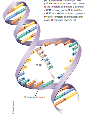
Protein synthesis actually takes place outside the cell nucleus, in the cytoplasm at the ribosomes. But the DNA molecule can’t leave the cell’s nucleus. Therefore the first step in protein synthesis is to copy the DNA message into a form of RNA called messenger RNA(mRNA), which can pass through the nuclear membrane into the cytoplasm. RNA is similar to DNA but it differs in some important ways:
- It’s single-stranded. (This is true for the forms we discuss here but not true for all forms of RNA.)
- It contains a different type of sugar.
- It contains the base uracil as a substitute for the DNA base thymine. (Uracil binds to adenine in the same way thymine does.)
The mRNA molecule forms on the DNA template in pretty much the same way that new DNA molecules do. As Table in DNA replication, the two DNA strands separate, but only partially, and one of these strands attracts free-floating RNA nucleotides (also produced in the cell), which are joined together on the DNA template. The formation of mRNA is called transcription because, in fact, the DNA code is being copied, or transcribed . Transcription continues until a section of DNA called a terminator region (composed of one of three specific DNA triplets) is reached and the process stops . At this point, the mRNA strand, comprising anywhere from 5,000 to perhaps as many as 200,000 nucleotides, peels away from the DNA model, and a portion of it travels through the nuclear membrane to the ribosome. Meanwhile, the bonds between the DNA bases are re-established and the DNA molecule is once more intact. As the mRNA strand arrives at the ribosome, its message is translated, or decoded. Just as each DNA triplet specifies one amino acid, so do mRNA triplets, which are called codons. Therefore the mRNA strand is “read” in codons, or groups of three mRNA bases at a time. Subsequently, another form of RNA, called transfer RNA (tRNA), brings each amino acid to the ribosome. The ribosome then joins that amino acid to another amino acid in the order dictated by the sequence of mRNA codons (or, ultimately, DNA triplets). In this way, amino acids are linked together to form a molecule that will eventually be a protein or part of a protein. But it’s important to mention that if a DNA base or sequence of bases is changed through mutation, some proteins may not be made or they may be defective. In this case, cells won’t function properly, or they may not function at all.