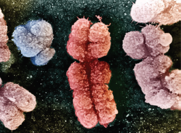
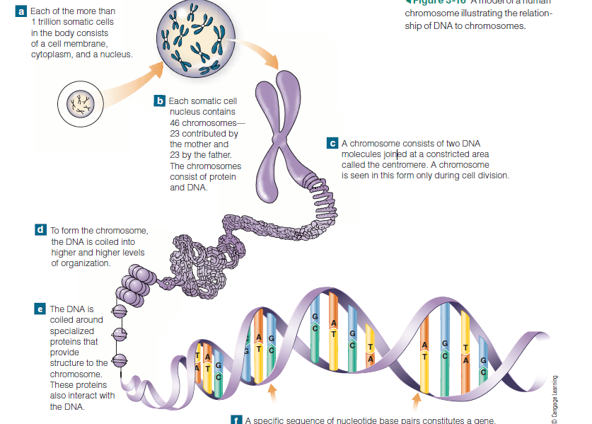
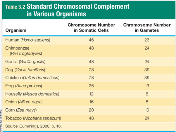
Throughout much of a cell’s life, its DNA (all 6 feet of it!) directs cellular functions and exists as an uncoiled, granular substance. However, at various times in the life of most types of cells, normal activities cease and the cell divides. Cell division produces new cells, and at the beginning of this process, the DNA becomes tightly coiled and is visible under a microscope as a set of discrete structures called chromosomes . Chromosomes are composed of a DNA molecule and proteins . During normal cell function, if the DNA were organized into chromosomes, they would be single stranded structures. However, during the early stages of cell division when they become visible, they’re made up of two strands, or two DNA molecules, joined together at a constricted area called the centromere. The reason there are two strands is simple: The DNA molecules have replicated, and one strand is an exact copy of the other. Every species has a specific number of chromosomes in somatic cells . Humans have 46, while chimpanzees and gorillas have 48. This point mutation a change in one of the four DNA bases. Chromosomes Discrete structures composed of DNA and proteins found only in the nuclei of cells. Chromosomes are visible under magnification only during certain phases of cell division doesn’t mean that humans have less DNA than chimpanzees and gorillas. It just means that the DNA is packaged differently.
There are two basic types of chromosomes: autosomes and sex chromosomes. Autosomes carry information that governs all physical characteristics except primary sex determination. The two sex chromosomes are the X and Y chromosomes; in mammals, the Y chromosome is directly involved in determining maleness. Although the X chromosome is called a sex chromosome, it actually functions more like an autosome.
Because it’s not involved in primary sex determination and it influences several other traits. Among mammals, all genetically normal females have two X chromosomes (XX), and they’re female only because they don’t have a Y chromosome. (Female is the default setting.) All genetically normal males have one X and one Y chromosome (XY). Chromosomes occur in pairs, so all normal human somatic cells have 22 pairs of autosomes and one pair of sex chromosomes (23 pairs in all). With few exceptions, abnormal numbers of autosomes are fatal—usually soon after conception. Although abnormal numbers of sex chromosomes aren’t usually fatal, they may result in sterility and frequently have other consequences. So to function normally, it’s essential for a human cell to possess both members of each chromosomal pair, or a total of 46 chromosomes.
Offspring inherit one member of each chromosomal pair from the father (the paternal chromosome) and one member from the mother (the maternal chromosome). Members of chromosomal pairs are alike in size and position of the centromere, and they carry genetic information governing the same traits. However, this doesn’t mean that partner chromosomes are genetically identical; it just means that they influence the same traits. For example, on both copies of a person’s ninth chromosome, there’s a locus, or gene position, that determines which of the four ABO blood types (A, B, AB, or O) he or she will have. However, these locus (pl., loci) (lo’-kus, lo-sigh’) the position or location on a chromosome where a given gene occurs. the term is sometimes used interchangeably with gene.
Two ninth chromosomes might not have identical DNA segments at the ABO locus. In other words, at numerous genetic loci, there may be more than one possible form of a gene, and these different forms are called alleles (Fig 3-17).
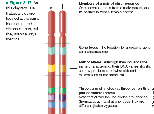
Alleles are alternate forms of a gene that can direct the cell to produce slightly different forms of a product, and ultimately, different expressions of a trait—as in the hemoglobin S (HbS) example. At the ABO locus, there are three possible alleles: A, B, and O. However, since individuals have only two ninth chromosomes, only two alleles are present in any one person. And the variation in alleles at the ABO locus is what accounts for the variation among humans in ABO blood type.
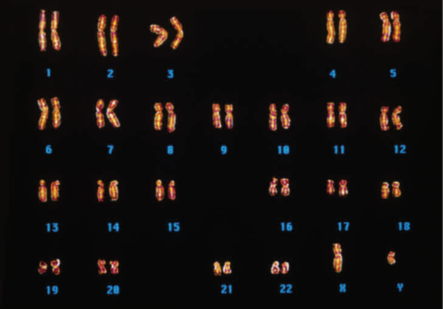
Karyotyping Chromosomes
One method frequently used to examine chromosomes in an individual is to produce a karyotype. The chromosomes used in karyotypes are obtained from dividing cells. White blood cells can be cultured, chemically treated, and microscopically examined to identify the ones that are dividing. These cells are then photographed through a microscope to produce photomicrographs of intact, double-stranded chromosomes. Partner chromosomes are then matched up, and the entire set is arranged in descending order by size so that the largest chromosome appears first.
Karyotyping has had numerous practical applications. Physicians and genetic counselors use karyotypes to help diagnose chromosomal disorders in patients, and they’re used in prenatal testing to identify chromosomal abnormalities in developing fetuses. Karyotype analysis has also revealed many chromosomal similarities shared by different species, including humans and nonhuman primates. But, now that scientists can directly compare the genomes of species, karyotyping probably won’t continue being used for this or several other purposes.